
Prior to Surgery of Ureteroscopic lithotripsy – Dr Vamsi’s Urology Hospital In Hyderabad
Preoperative Testing
During your PMC visit for Ureteroscopic lithotripsy Treatment, the items listed below will be ordered as deemed necessary based upon your age and medical history.
You will have an opportunity to speak with the anaesthesia staff regarding the types of anaesthesia available and their relative risks and benefits.
- EKG (electrocardiogram)
- CBC (complete blood count)
- PT / PTT (blood coagulation profile)
- Comprehensive Metabolic Panel (blood chemistry profile)
- Urinalysis
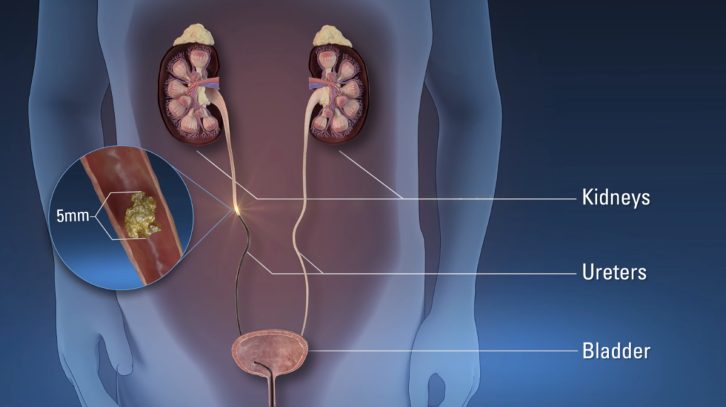
Lithotripsy is a medical procedure that uses shock waves or a laser to break down stones in the kidney, gallbladder, or ureter. The remaining particles of small stone will exit the body when a person urinates.
Diet Day Before Surgery
You may eat a regular diet until midnight the night before surgery. After midnight please do not eat or drink anything. If instructed to do so, you may take your prescription medications with a sip of water.
Surgery Procedure
Once asleep a small instrument (ureteroscope) is passed through the urethra (water channel) and bladder into the ureter (tube transporting urine from the kidney to the bladder).
The Ureteroscopic lithotripsy contains a light and camera that projects an image onto a monitor next to the operating room table.
The scope is advanced until the stone is located. If the stone is small it may be grabbed with a basket device and removed intact. However, in most instances, the stone will be too big to be safely removed whole.
If so, a laser will be used to break the stone up into several small pieces (lithotripsy) that are grabbed and removed.
Alternatively, the laser may be used to turn the stone into numerous small fragments that drain out of the kidney on their own following surgery.
In most cases, a urethral stent (a piece of surgical plastic that goes from the kidney to the bladder through the ureter) will be placed at the end of the procedure.
The stent keeps the ureter open following surgery. If a stent is not placed the ureter may temporarily swell shut or become occluded by blood clots or stone debris resulting in kidney pain following surgery.
In a small number of cases (less than 5%), the ureter is too narrow to safely permit passage of the ureteroscopy into it. When this occurs a urethral stent is placed and stone removal is delayed one to two weeks. The stent gently stretches the ureter allowing safe passage of the scope at a later date.